Saturday, December 4, 2010

Tongue


The tongue is a muscular hydrostat on the floors of the mouths of most vertebrates which manipulates foodmastication. It is the primary organ of taste, as much of the upper surface of the tongue is covered in papillae and taste buds. It is sensitive and kept moist by saliva, and is richly supplied with nerves and blood vessels. In humans a secondary function of the tongue is phonetic articulation. The tongue also serves as a natural means of cleaning one's teeth. for

Musculature

The eight muscles of the human tongue are classified as either intrinsic or extrinsic. The four intrinsic muscles act to change the shape of the tongue, and are not attached to any bone. The four extrinsic muscles act to change the position the tongue, and are anchored to bone.

 Intrinsic muscles

  1. Superior longitudinal fibers: shorten the tongue.
  2. Inferior longitudinal fibers: shorten the tongue.
  3. Vertical fibers: flatten and widen the tongue.
  4. Transverse fibers: narrow and elongate the tongue.

 Extrinsic muscles

  1. Genioglossus
  2. Hyoglossus
  3. Styloglossus
  4. Palatoglossus

The receptors for taste, called taste  buds,are situated chiefly in the tongue, but they are also located in the roof of the mouth and near the pharynx. They are able to detect four basic tastes: salty, sweet, bitter, and sour. The tongue also can detect a sensation called "umami" from taste receptors sensitive to amino acids. Generally, the taste buds close to the tip of the tongue are sensitive to sweet tastes, whereas those in the back of the tongue are sensitive to bitter tastes. The taste buds on top and on the side of the tongue are sensitive to salty and sour tastes. At the base of each taste bud there is a nerve that sends the sensations to the brain. The sense of taste functions in coordination with the sense of smell. The number of taste buds varies substantially from individual to individual, but greater numbers increase sensitivity. Women, in general, have a greater number of taste buds than men. As in the case of color blindness, some people are insensitive to some tastes.
 
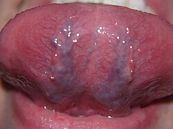
The underside of a human tongue,
The tongue receives its blood supply primarily from the lingual artery, a branch of the external carotid artery. The floor of the mouth also receives its blood supply from the lingual artery. The triangle formed by the intermediate tendon of the digastric muscle, the posterior border of the mylohyoid muscle, and the hypoglossalPirogov's, Pirogoff's, or Pirogov-Belclard's triangle. The lingual artery is a good place to stop severe hemorrage from the tongue. nerve is sometimes called
There is also secondary blood supply to the tongue from the tonsillar branch of the facial artery and the ascending pharyngeal artery.


Nerve supply

Taste for the anterior 2/3 of the tongue is supplied by the Facial nerve (Chorda tympani, CN7). General sensation of the anterior 2/3 is supplied by the Lingual nerve which is a branch of V3 of the Trigeminal nerve CN V.
Taste as well as general sensation for the posterior 1/3 is supplied by the Glossopharyngeal nerve (CN 9).
All intrinsic and extrinsic muscles of the tongue are supplied by the Hypoglossal nerve (CN 12), except for one of the extinsic muscles, palatoglossus, which is inervated by CN10 of the pharyngeal plexus.


Length

The average length of the human tongue from the oropharynx to the tip is 10 cm .

Monday, November 29, 2010

Lips

Lips are a visible body part at the mouth of humans and many animals. Lips are soft, movable, and serve as the opening for food intake and in the articulation of sound and speech. Human lips are a tactile sensory organ, and can be erogenous when used in kissing and other acts of intimacy.

Anatomical basics of the human lip

One differentiates between the upper (Labium superius) and lower lip (Labium inferius). The lower lip is usually somewhat larger. The border between the lips and the surrounding skin is referred to as the vermillion border, or simply the vermilion. The vertical groove on the upper lip is known as the philtrum.
The skin of the lip, with three to five cellular layers, is very thin compared to typical face skin, which has up to 16 layers. With light skin color, the lip skin contains fewer melanocytes (cells which produce melanin pigment , which give skin its color). Because of this, the blood vessels appear through the skin of the lips, which leads to their notable red coloring. With darker skin color this effect is less prominent, as in this case the skin of the lips contains more melanin and thus is visually darker. The skin of the lip forms the border between the exterior skin of the face, and the interior mucous membrane of the inside of the mouth.
The lip skin is not hairy, and does not have sweat glands or sebaceous glands. Therefore it does not have the usual protection layer of sweat and body oils which keep the skin smooth, inhibit pathogens, and regulate warmth. For these reasons, the lips dry out faster and become chapped more easily.

Eye


The eye is the organ of vision. It has a complex structure consisting of a transparent lens that focuses light on the retina. The retina is covered with two basic types of light-sensitive cells-rods and cones. The cone cells are sensitive to color and are located in the part of the retina called the fovea, where the light is focused by the lens. The rod cells are not sensitive to color, but have greater sensitivity to light than the cone cells. These cells are located around the fovea and are responsible for peripheral vision and night vision. The eye is connected to the brain through the optic nerve. The point of this connection is called the "blind spot" because it is insensitive to light. Experiments have shown that the back of the brain maps the visual input from the eyes.
The brain combines the input of our two eyes into a single three-dimensional image. In addition, even though the image on the retina is upside-down because of the focusing action of the lens, the brain compensates and provides the right-side-up perception. Experiments have been done with subjects fitted with prisms that invert the images. The subjects go through an initial period of great confusion, but subsequently they perceive the images as right side up.


The range of perception of the eye is phenomenal. In the dark, a substance produced by the rod cells increases the sensitivity of the eye so that it is possible to detect very dim light. In strong light, the iris contracts reducing the size of the aperture that admits light into the eye and a protective obscure substance reduces the exposure of the light-sensitive cells. The spectrum of light to which the eye is sensitive varies from the red to the violet. Lower electromagnetic frequencies in the infrared are sensed as heat, but cannot be seen. Higher frequencies in the ultraviolet and beyond cannot be seen either, but can be sensed as tingling of the skin or eyes depending on the frequency. The human eye is not sensitive to the polarization of light, i.e., light that oscillates on a specific plane. Bees, on the other hand, are sensitive to polarized light, and have a visual range that extends into the ultraviolet. Some kinds of snakes have special infrared sensors that enable them to hunt in absolute darkness using only the heat emitted by their prey. Birds have a higher density of light-sensing cells
than humans do in their retinas, and therefore, higher visual acuity.


Color blindness or "Dalton-ism" is a common abnormality in human vision that makes it impossible to differentiate colors accurately. One type of color blindness results in the inability to distinguish red from green. This can be a real handicap for certain types of occupations. To a colorblind person, a person with normal color vision would appear to have extrasensory perception. However, we want to reserve the term "extrasensory perception" for perception that is beyond the range of the normal.


Eyes are organs that detect light, and convert it to electro-chemical impulses in neurons. The simplest photoreceptors in conscious vision connect light to movement. In higher organisms the eye is a complex optical system which collects light from the surrounding environment; regulates its intensity through a diaphragm; focuses it through an adjustable assembly of lenses to form an image; converts this image into a set of electrical signals; and transmits these signals to the brain, through complex neural pathways that connect the eye, via the optic nerve, to the visual cortex and other areas of the
brain. Eyes with resolving power have come in ten fundamentally different forms, and 96% of animal species possess a complex optical system.[1] Image-resolving eyes are present in molluscs, chordates and arthropods.[2]
The simplest "eyes", such as those in microorganisms, do nothing but detect whether the surroundings are light or dark, which is sufficient for the entrainment of circadian rhythms. From more complex eyes, retinal photosensitive ganglion cells send signals along the retinohypothalamic tract to the suprachiasmatic nuclei to effect circadian adjustment.

Kidney

The kidneys are two organs with several functions. They are seen in many types of animals, including vertebrates and some invertebrates. They are an essential part of the urinary system and also serve homeostatic functions such as the regulation of electrolytes, maintenance of acid-base balance, and regulation of blood pressure. They serve the body as a natural filter of the blood, and remove wastes which are diverted to the urinary bladder. In producing urine, the kidneys excrete wastes such as urea and ammonium; the kidneys also are responsible for the reabsorption of water, glucose, and amino acids. The kidneys also produce hormones including calcitriol, renin, and erythropoietin.
Located at the rear of the abdominal cavity in the retroperitoneum, the kidneys receive blood from the paired renal arteries, and drain into the paired renal veins. Each kidney excretes urine into a ureter, itself a paired structure that empties into the urinary bladder.
Renal physiology is the study of kidney function, while nephrology is the medical specialty concerned with kidney diseases. Diseases of the kidney are diverse, but individuals with kidney disease frequently display characteristic clinical features. Common clinical conditions involving the kidney include the nephritic and nephrotic syndromes, renal cysts, acute kidney injury, chronic kidney disease, urinary tract infection, nephrolithiasis, and urinary tract obstruction.[1] Various cancers of the kidney exist; the most common adult renal cancer is renal cell carcinoma. Cancers, cysts, and some other renal conditions can be managed with removal of the kidney, or nephrectomy. When renal function, measured by glomerular filtration rate, is persistently poor, dialysis and kidney transplantation may be treatment options. Although they are not severely harmful, kidney stones can be a pain and a nuisance. The removal of kidney stones includes sound wave treatment, which breaks up the stones into smaller pieces which are then passed through the urinary tract. One common symptom of kidney stones is a sharp pain in the medial/lateral segments of the lower back.

Nose

 
The nose is the organ responsible for the sense of smell. The cavity of the nose is lined with mucous membranes that have smell receptors connected to the olfactory nerve. The smells themselves consist of vapors of various substances. The smell receptors interact with the molecules of these vapors and transmit the sensations to the brain. The nose also has a structure called the vomeronasal organ whose function has not been determined, but which is suspected of being sensitive to pheromones that influence the reproductive cycle. The smell receptors are sensitive to seven types of sensations that can be characterized as camphor, musk, flower, mint, ether, acrid, or putrid. The sense of smell is sometimes temporarily lost when a person has a cold. Dogs have a sense of smell that is many times more sensitive than man's.


Physically a nose is an organ on the face. Anatomically, a nose is a protuberance in vertebrates that houses the nostrils, or nares, which admit and expel air for respiration in conjunction with the mouth. Behind the nose is the olfactory mucosa and the sinuses. Behind the nasal cavity, air next passes through the pharynx, shared with the digestive system, and then into the rest of the respiratory system. In humans, the nose is located centrally on the face; on most other mammals, it is on the upper tip of the snout.

Air conditioning


As an interface between the body and the external world, the nose and associated structures frequently perform additional functions concerned with conditioning entering air (for instance, by warming and/or humidifying it, also for flicking if moving and by mostly reclaiming moisture from the air before it is exhaled (as occurs most efficiently in camels). The nose often has inner hairs whose function is to stop unwanted particles from entering the lungs.

Sense of direction

The wet nose of dogs is useful for the perception of direction. The sensitive cold receptors in the skin detect the place where the nose is cooled the most and this is the direction a particular smell that the animal just picked up comes from.[1]

Mouth

The mouth is the first portion of the alimentary canal that receives food and saliva.The oral mucosa is the mucous membrane epithelium lining the inside of the mouth.
In addition to its primary role as the beginning of the digestive system, in humans the mouth also plays a significant role in communication. While primary aspects of the voice are produced in the throat, the tongue, lips, and jaw are also needed to produce the range of sounds included in human language. Another non-digestive function of the mouth is its role in secondary social and/or sexual activity, such as kissing.
The mouth is normally moist, and is lined with a mucous membrane. The lips mark the transition from mucous membrane to skin, which covers most of the body.


Mouth cavity

The first space of the mouth is the mouth cavity, bounded laterally and in front by the alveolar arches (containing the teeth), and posteriorily by the isthmus of the fauces. The oral cavity is also known as the mouth which swallows food and drinks that then go down the esophagus and into the stomach.

Function

The mouth plays an important role in sucking (it is part of the sucking apparatus), facial expression, eating, drinking and breathing. Infants are born with a sucking reflex, by which they instinctively know to suck for nourishment using their lips and jaw.

Cultural aspects

According to western etiquette, the mouth is kept closed, especially when chewing.
Lips can be adorned with lipstick or lip gloss, although in most cultures this is typically only practiced by females. Both men and women, however, apply lip balm in order to soothe chapped or dry lips.
Piercings in or around the mouth have been made popular by younger generations, including those on the lip or tongue. The uvula piercing, while increasing in popularity, remains relatively rare.

Development

The philtrum is the vertical groove in the upper lip, formed where the nasomedial and maxillary processes meet during embryo development. When these processes fail to fuse fully, a hare lip and/or cleft palate can result.
The nasolabial folds are the deep creases of tissue that extend from the nose to the sides of the mouth. One of the first signs of age on the human face is the increase in prominence of the nasolabial folds.

Ear

 
The ear is the organ of hearing. The outer ear protrudes away from the head and is shaped like a cup to direct sounds toward the tympanic membrane, which transmits vibrations to the inner ear through a series of small bones in the middle ear called the malleus, incus and stapes. The inner ear, or cochlea, is a spiral-shaped chamber covered internally by nerve fibers that react to the vibrations and transmit impulses to the brain via the auditory nerve. The brain combines the input of our two ears to determine the direction and distance of sounds.


The inner ear has a vestibular system formed by three semicircular canals that are approximately at right angles to each other and which are responsible for the sense of balance and spatial orientation. The inner ear has chambers filled with a viscous fluid and small particles (otoliths) containing calcium carbonate. The movement of these particles over small hair cells in the inner ear sends signals to the brain that are interpreted as motion and acceleration. 


The human ear can perceive frequencies from 16 cycles per second, which is a very deep bass, to 28,000 cycles per second, which is a very high pitch. Bats and dolphins can detect frequencies higher than 100,000 cycles per second. The human ear can detect pitch changes as small as 3 hundredths of one percent of the original frequency in some frequency ranges. Some people have "perfect pitch", which is the ability to map a tone precisely on the musical scale without reference to an external standard. It is estimated that less than one in ten thousand people have perfect pitch, but speakers of tonal languages like Vietnamese and Mandarin show remarkably precise absolute pitch in reading out lists of words because pitch is an essential feature in conveying the meaning of words in tone languages. The Eguchi Method teaches perfect pitch to children starting before they are 4 years old. After age 7, the ability to recognize notes does not improve much.


The ear is the anatomical organ that detects sound. It not only acts as a receiver for sound, but also plays a major role in the sense of balance and body position. The ear is part of the auditory system.
The word "ear" may be used correctly to describe the entire organ or just the visible portion. In most mammals, the visible ear is a flap of tissue that is also called the pinna and is the first of many steps in hearing. In people, the pinna is often called the auricle. Vertebrates have a pair of ears, placed symmetrically on opposite sides of the head. This arrangement aids in the ability to localize sound sources.

Introduction to ears and hearing


Audition is the scientific name for the sense of sound. Sound is a form of energy that moves through air, water, and other matter, in waves of pressure. Sound is the means of auditory communication, including frog calls, bird songs and spoken language. Although the ear is the vertebrate sense organ that recognizes sound, it is the brain and central nervous system that "hears". Sound waves are perceived by the brain through the firing of nerve cells in the auditory portion of the central nervous system. The ear changes sound pressure waves from the outside world into a signal of nerve impulses sent to the brain.

ntroduction to ears and hearing

Audition is the scientific name for the sense of sound. Sound is a form of energy that moves through air, water, and other matter, in waves of pressure. Sound is the means of auditory communication, including frog calls, bird songs and spoken language. Although the ear is the vertebrate sense organ that recognizes sound, it is the brain and central nervous system that "hears". Sound waves are perceived by the brain through the firing of nerve cells in the auditory portion of the central nervous system. The ear changes sound pressure waves from the outside world into a signal of nerve impulses sent to the brain.The outer part of the ear collects sound. That sound pressure is amplified through the middle portion of the ear and, in land animals, passed from the medium of air into a liquid medium. The change from air to liquid occurs because air surrounds the head and is contained in the ear canal and middle ear, but not in the inner ear. The inner ear is hollow, embedded in the temporal bone, the densest bone of the body. The hollow channels of the inner ear are filled with liquid, and contain a sensory epithelium that is studded with hair cells. The microscopic "hairs" of these cells are structural protein filaments that project out into the fluid. The hair cells are mechanoreceptors that release a chemical neurotransmitter when stimulated. Sound waves moving through fluid push the filaments; if the filaments bend over enough it causes the hair cells to fire. In this way sound waves are transformed into nerve impulses. In vision, the rods and cones of the retina play a similar role with light as the hair cells do with sound. The nerve impulses travel from the left and right ears through the eighth cranial nerve to both sides of the brain stem and up to the portion of the cerebral cortex dedicated to sound. This auditory part of the cerebral cortex is in the temporal lobe.